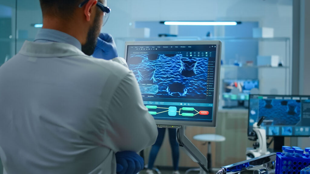
Medical imaging has long been a crucial part of contemporary healthcare, allowing for the visualization and investigation of the interior organs and bodily processes. These imaging technologies, which include X-rays, MRI, and CT scans, have changed diagnosis, planning of the patient’s course of treatment, and patient health monitoring.
However, the demand for cutting-edge technologies to interpret and handle the enormous quantity of data created by these systems has been driven by the quest for more accurate, quicker, and more thorough medical imaging. Field-Programmable Gate Arrays, or FPGAs, have become crucial instruments in the field of medical imaging as a result of this effort.
FPGA Applications in Medical Imaging
Due to its high performance, real-time processing, and flexibility, Field-Programmable Gate Arrays (FPGAs) have a wide range of applications in the field of medical imaging. The following are some crucial areas where FPGAs are essential to the advancement of medical imaging:
A. Preprocessing and Data Acquisition
Image Enhancement: FPGAs play a key role in improving the caliber of medical imaging. They can be designed to use different noise reduction, contrast enhancement, and edge detection filters and algorithms. FPGAs aid in enhancing image clarity and lowering radiation exposure in real-time imaging, such as X-ray fluoroscopy.
Noise Reduction: Data capture noise is a common problem for medical pictures. In order to maintain the clarity and accuracy of the diagnostic data, FPGAs can incorporate cutting-edge noise reduction techniques like adaptive filtering and wavelet denoising.
B. Image Reconstruction and Processing
CT and MRI Image Reconstruction: For quick image reconstruction in computed tomography (CT) and magnetic resonance imaging (MRI), FPGAs are widely used. They shorten the time patients spend in scanners and enable real-time interventional operations by speeding up the intricate mathematical transformations required to turn raw data into detailed 2D or 3D images.
Ultrasound Beamforming: FPGA-based beamforming is essential for producing high-quality images in ultrasonic imaging. Using massively parallel computing, FPGAs focus the ultrasound beam to create exact images of tissues and organs by combining inputs from several transducer elements.
C. Real-Time Image Analysis
Detection of Anomalies and Abnormalities: FPGAs make it possible to analyze medical images in real time and detect anomalies and abnormalities automatically. For instance, in mammography, FPGAs might help radiologists by pointing out problematic spots that would need additional inspection, potentially assisting in the early diagnosis of cancer.
Motion Tracking and Registration: FPGAs are used for real-time motion tracking and picture registration during processes like image-guided surgery. This improves surgical accuracy and lowers patient risk by ensuring that surgical tools are properly guided depending on the anatomy of the patient.
FPGA Design Considerations for Medical Imaging
When integrating these reconfigurable components into medical imaging systems, effective FPGA design is essential. To successfully integrate FPGAs for activities ranging from image processing to real-time analysis, designers must manage a wide range of hardware and software considerations. In this section, we look into the crucial factors to take into account when creating FPGA-based medical imaging solutions:
A. Hardware and Software Development Tools
Selection of FPGA Platform: A crucial choice is which FPGA platform to choose. It entails taking into account elements including the FPGA’s processing capability, resources, and suitability for jobs involving medical imaging. Xilinx and Intel (formerly Altera), two common FPGA providers, each offer a variety of devices with different capabilities.
Programming Language and Tools: Deciding on the programming language and development tools is essential. VHDL and Verilog are popular hardware description languages for FPGA development. Additionally, specialized development environments like Xilinx Vivado and Intel Quartus Prime simplify FPGA programming and synthesis.
B. Resource Management and Optimization
Resource Utilization: Maximizing FPGA resource utilization is vital to efficiently perform medical image processing. Designers must carefully manage the allocation of logic elements, memory, and DSP blocks to ensure optimal use of the FPGA’s resources.
Power Efficiency: Power consumption is a critical concern, particularly in portable medical devices. FPGA designs should aim to minimize power usage while maintaining high performance. Techniques such as clock gating and low-power design methodologies are essential in this context.
C. Safety and Reliability Concerns
Redundancy and Fault Tolerance: In medical imaging applications, reliability is paramount. Designers should implement redundancy and fault tolerance mechanisms to ensure uninterrupted operation, especially in critical diagnostic and monitoring systems.
Testing and Verification: Rigorous testing and verification procedures are necessary to identify and rectify potential design flaws. Techniques like simulation, formal verification, and hardware-in-the-loop testing are essential to validate FPGA-based medical imaging systems.
D. Compliance with Regulatory Standards
Medical Device Regulations: Medical imaging devices are subject to stringent regulatory requirements to ensure patient safety and data integrity. FPGA-based systems must comply with standards such as ISO 13485, IEC 62304, and FDA guidelines to facilitate approval and market entry.
Data Security and Privacy: Protection of patient data and compliance with data privacy regulations, such as HIPAA in the United States, are non-negotiable. FPGA designs should incorporate encryption and secure data handling protocols to safeguard sensitive medical information.
Challenges and Ethical Considerations
While FPGAs offer significant advantages in the field of medical imaging, they also present various challenges and raise ethical considerations that must be addressed to ensure their responsible and effective use.
A. Privacy and Security Issues in Medical Imaging
Data Security: Medical picture transmission and storage must be protected from unauthorized access and potential breaches since they frequently contain sensitive patient data. Real-time data processing using FPGAs calls for strong access control and encryption protocols.
Cybersecurity: FPGAs are vulnerable to cybersecurity threats, including malware and hardware attacks. Ensuring the security of FPGA-based medical imaging systems is paramount to prevent data tampering or system compromise.
B. Ethical Use of Medical Data
Informed Consent: Strict informed consent guidelines must be followed when collecting and using medical imaging data for study or diagnosis. Patients should be fully informed about how and why their data will be utilized.
Data Ownership: Determining the ownership of medical imaging data, especially when it is used for research or training AI models, can be a complex issue. Ethical frameworks need to address who has the right to use and benefit from such data.
C. Regulatory and Legal Framework
Compliance: FPGAs used in medical imaging must adhere to legal requirements such as the General Data Protection Regulation (GDPR) in the European Union or the Health Insurance Portability and Accountability Act (HIPAA) in the United States. To prevent legal repercussions, compliance with these requirements must be guaranteed.
Liability: Liability issues arise from the usage of FPGAs in medical imaging in the event of mistakes or malfunctions. For patient safety and legal remedies, defining precise legal obligations and accountability for FPGA-based systems is essential.
D. Ethical Considerations in AI Integration
Bias and Fairness: When FPGAs are integrated with AI algorithms for image analysis, ethical concerns related to bias and fairness in AI models become pertinent. Efforts must be made to reduce bias and ensure equitable healthcare outcomes.
Transparency: Ensuring transparency in AI decision-making processes is crucial. Healthcare practitioners should be able to understand and interpret AI-driven diagnostic recommendations to maintain the human touch in patient care.
Conclusion
FPGAs have completely changed the way that medical imaging is done in the healthcare industry. The adaptability and real-time processing capacity needed to improve diagnostic precision and therapeutic efficacy are provided by FPGAs.
In conclusion, FPGAs have evolved into essential tools for addressing the changing requirements of contemporary medical imaging, enabling quicker, more precise diagnosis and treatment planning. Their flexibility and processing power have the potential to transform healthcare even further, bringing technology and medicine closer together in a fascinating way.


![What is FPGA Introduction to FPGA Basics [2023] computer-chip-dark-background-with-word-intel-it](https://fpgainsights.com/wp-content/uploads/2023/06/computer-chip-dark-background-with-word-intel-it-300x171.jpg)


